
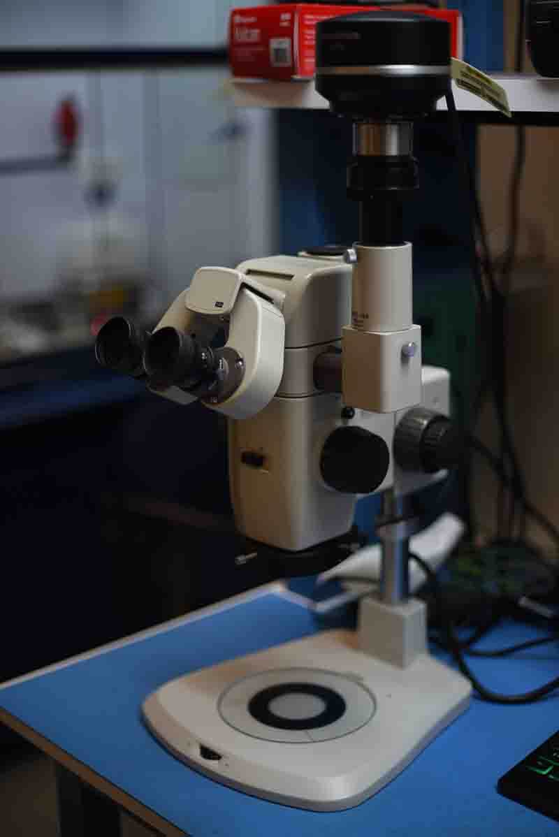
To perform low power optical microscopy, we utilize a Nikon SMZ stereo zoom optical microscope for its excellent low magnification optical technology. There are a number of attributes which make this microscope a foundational tool to labs across multiple industries. The long working distance realized by the objective lens permits samples to be manipulated and worked under the microscope in a magnified environment. The wide field of view allows the analyst to simultaneously inspect a large sample area without the need to move or disrupt the sample. Thanks in part to the long focal length and clear optical glass used in the objectives, this system renders images which are crisp and exhibit an impressive perception of depth.
The applications this system was designed to serve are diverse:
Advance Imaging
For more information and to learn more about this microscope visit: NikonSMZ
The microscope is topped off with the Olympus DP72 12.2 mega pixel digital camera. This camera was designed and manufactured by Olympus for high resolution, high color production, and high sensitivity. By shifting the imaging sensor in sub pixel steps this camera is capable of creating superior quality images with nine times the details of stationary cameras. The Peltier cooled imaging sensor allows the camera to capture images under low light conditions such as dark field and fluorescence images.
While this camera has been discontinued and replaced by the DP74, information can be found at the following link: DP72
Capable of 7x to 63x magnification under bright field lighting for imaging. Image capture up to 12 megapixel and calibrated measurements. Typically used to capture images of electronic package, electronic components, medical devices, and printed circuit boards.
Image capture and post image analysis is performed with Olympus Stream software. Olympus Stream allows us to perform calibrated measurements, quantitative analysis, and reporting. For more information on Olympus Stream image capture software visit: Stream
The applications this system was designed to serve are diverse:
Advance Imaging
- Count and Measure Solution
- Three-dimensional image display and measurement
- Metallurgical inspection for nodularity, grain sizing, and flake analysis
- Grain sizing using the intercept counting method
- Grain sizing using the planimetric method
- Graphite nodularity evaluation
- Rating the non-metallic inclusion content
- Comparison of sample images with reference standards
- Dendrite arm spacing measurement
- Welding distortion
- Phase and region of interest measurements
- Particle distribution
- Throwing power management
- Automatic critical dimension measurements
- Thin coating thickness evaluation (calotest method)
- Layer thickness measurement
- Pore fraction and density measurement
- Surface or external examination for incoming quality inspection
- Examining composites, Fabrics, textiles, and asbestos
- Examination and analysis of Optoelectronics
- Examination and analysis of micro electro mechanical systems (MEMs)
- Examination and analysis of microelectronic packaging and devices
- Inspection of PCB assemblies
- Examination and analysis of implants and protheses
- General photo documentation
For more information and to learn more about this microscope visit: NikonSMZ
The microscope is topped off with the Olympus DP72 12.2 mega pixel digital camera. This camera was designed and manufactured by Olympus for high resolution, high color production, and high sensitivity. By shifting the imaging sensor in sub pixel steps this camera is capable of creating superior quality images with nine times the details of stationary cameras. The Peltier cooled imaging sensor allows the camera to capture images under low light conditions such as dark field and fluorescence images.
While this camera has been discontinued and replaced by the DP74, information can be found at the following link: DP72
Capable of 7x to 63x magnification under bright field lighting for imaging. Image capture up to 12 megapixel and calibrated measurements. Typically used to capture images of electronic package, electronic components, medical devices, and printed circuit boards.
Image capture and post image analysis is performed with Olympus Stream software. Olympus Stream allows us to perform calibrated measurements, quantitative analysis, and reporting. For more information on Olympus Stream image capture software visit: Stream